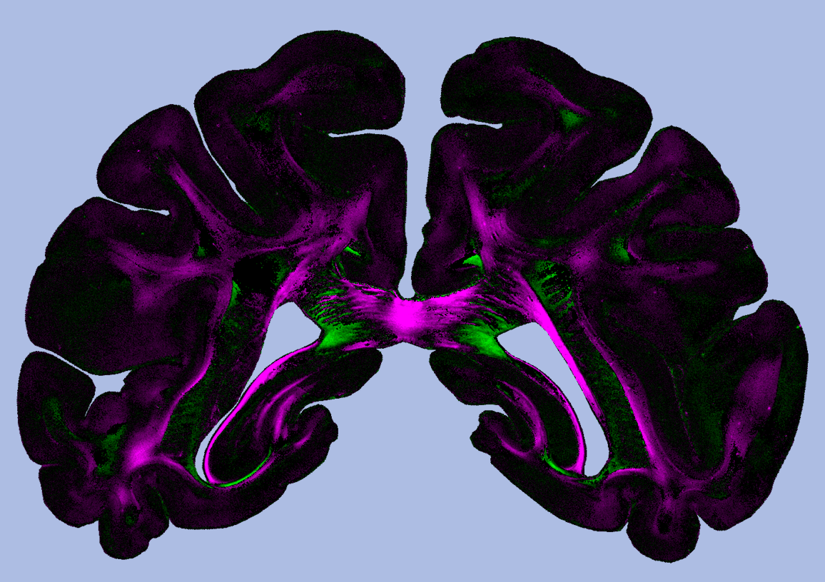
Diattenuation Imaging – A promising imaging technique for brain research
March 19, 2019: We developed Diattenuation Imaging, a new imaging technique that provides structural brain tissue information that was previously difficult to access.
Diattenuation determines how much the intensity of polarized light is reduced when it passes through a brain section. We found that in some brain regions, the tissue is maximally transparent when the polarization of the light is oriented parallel to the nerve ffibersibres. In other brain regions, the tissue is maximally transparent when the polarization is oriented perpendicularly to the nerve fibers. The oberserved effects depend on tissue properties like the diameter of the fibers or the thickness of the surrounding myelin sheaths. This allows to use Diattenuation Imaging in order to differentiate, e.g., regions with many thin nerve fibers from regions with few thick nerve fibers. With current imaging methods, these tissue types cannot easily be distinguished. In addition, the new technology helps to make pathological changes visible and to identify connected regions and tissue types, assisting the complex reconstruction of the brain.
Original Publication: Menzel M, Axer M, Amunts K, De Raedt H, Michielsen K. “Diattenuation Imaging reveals different brain tissue properties”. Sci Rep. 2019;9:1939
