Publication in Nature Communications
November 5, 2025: We have published a paper in Nature Communications about the ability of Computational Scattered Light Imaging to visualize directed structures in all kinds of histological tissue samples, independent of sample preparation. This study has been made possible by a collaboration between Stanford University (USA), Forschungszentrum Jülich (Germany), and Erasmus MC in Rotterdam (Netherlands).
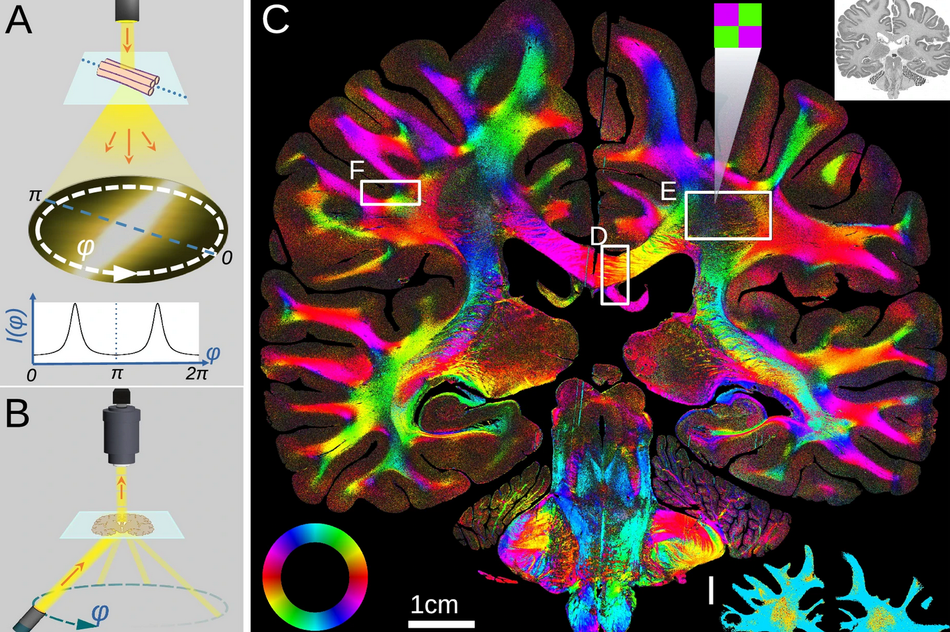
Mapping the brain’s fiber network is crucial for understanding its function and malfunction, but resolving nerve trajectories over large fields of view is challenging. Here, we show that computational scattered light imaging (ComSLI) can map fiber networks in histology independent of sample preparation, also in formalin-fixed paraffin-embedded (FFPE) tissues including whole human brain sections. We showcase this method in new and archived, animal and human brain sections, for different sample preparations (in paraffin, deparaffinized, various stains, unstained fresh-frozen). We convert microscopic orientations to microstructure-informed fiber orientation distributions (μFODs). Adapting tractography tools from diffusion magnetic resonance imaging (dMRI), we trace axonal trajectories revealing white and gray matter connectivity. These allow us to identify altered microstructure or deficient tracts in demyelinating or neurodegenerating pathology, and to show key advantages over dMRI, polarization microscopy, and structure tensor analysis. Finally, we map fibers in non-brain tissues, including muscle, bone, and blood vessels, unveiling the tissue’s function. Our cost-effective, versatile approach enables micron-resolution studies of intricate fiber networks across tissues, species, diseases, and sample preparations, offering new dimensions to neuroscientific and biomedical research.