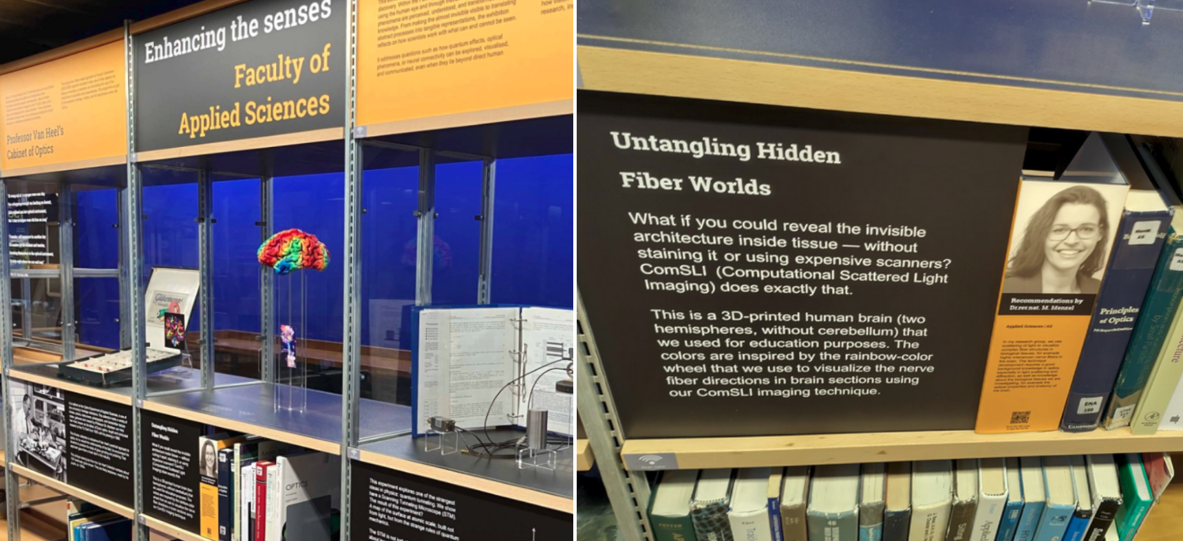
SPIE Photonics Europe Presentations
April 16, 2026: Hamed Abbasi, Mark Vermeulen, and Loes Ettema have presented their work at SPIE Photonics Europe in Strasbourg, France. Hamed talked about “Multimodal computational scattered light microscopy for collagen-specific whole-slide fiber mapping”, Mark about “Quantitative 3D-orientation mapping of biological fibers in computational scattered light imaging enabled through 3D-nanoprinted phantoms”, and Loes presented a poster on “Revealing fiber orientations on biological tissue surfaces with backscattered light imaging”.

Multimodal computational scattered light microscopy for collagen-specific whole-slide fiber mapping
The organization of collagen fibers in the extracellular matrix provides valuable insights into tumor prognosis for many solid tumors, as cancer cells and collagen fibers interact dynamically during tumor progression. Mapping peritumoral collagen fiber directionality can therefore enable more personalized prognoses, leading to improved treatment strategies and better patient outcomes. Computational Scattered Light Imaging (ComSLI) is a novel whole-slide microscopy technique that leverages anisotropic light scattering to reveal microscopic fiber orientations in any histology section. However, it lacks specificity for collagen, as other fiber types (e.g., muscle or nerve) are also visualized. In this study, we integrated crossed-polarizers, and excitation and emission filters for fluorescence microscopy into the optical path of the microscope. This modification enables us to visualize collagen in picrosirius red- and hematoxylin and eosin-stained microscopy slides, allowing for the generation of collagen-specific fiber orientation maps.
Quantitative 3D-orientation mapping of biological fibers in computational scattered light imaging enabled through 3D-nanoprinted phantoms
Computational Scattered Light Imaging (ComSLI) images the orientations of complex biological fiber structures with micrometer resolution and a large field of view. ComSLI works on a wide range of samples, including nerve, muscle, collagen, stained, unstained and non-birefringent tissues. It requires only an LED light source and camera, making it attractive for histological tissue analysis. While previous research observed an effect of out-of-plane fiber inclination on the ComSLI scattering patterns, a quantitative description has not been possible until now due to a lack of a priori knowledge of the inclination angle. Here, we performed measurements on 3D-nanoprinted fiber bundles with different inclination angles. We were able to show that the curvature observed in the measured scattering patterns matches conical diffraction by an inclined grating, allowing for a quantitative determination of the fiber inclination. The findings were validated on brain fiber bundles with known fiber inclination angles.
Revealing fiber orientations on biological tissue surfaces with backscattered light imaging
Visualizing fiber network organization in biological tissues is important for studying the relation between fiber organization and tissue functioning. With computational scattered light imaging (ComSLI), individual fiber bundle orientations can be resolved at micrometer resolution, even in densely interwoven networks. Up to now, the technique has mostly been used for visualizing fiber networks in thin tissue slices. Here, we present the use of ComSLI in backscattering mode for enabling measurements on biological tissue samples without the necessity of tissue slicing. We present recent findings on various tissues, including brain samples from different species, investigate the ability to extract crossing fibers, and study the effects of different wavelengths. Facilitating backscattered ComSLI measurements simplifies the sample preparation process and might open up new applications like intraoperative tissue assessment.