Bachelor & Master Thesis Projects
We welcome driven students with a background in applied physics, nanobiology, microscopy, data science, or similar who want to work at the interface of computational imaging and biomedical research - either on the experimental side (microscopy, hardware, tissue preparation) or on the computational side (image processing, signal analysis, simulations).
If you’re interested in a Master thesis project, please contact Miriam Menzel for possible projects (see below for examples of possible theses).
We have currently only Master thesis projects available.
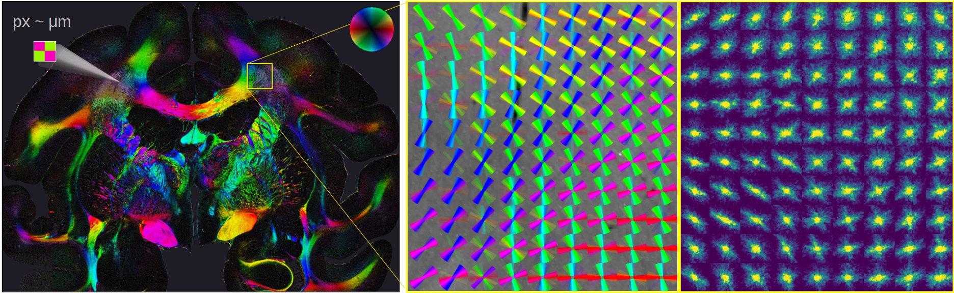
Computational Scattered Light Imaging (ComSLI) resolves fiber pathways (like nerve, muscle, or collagen fibers) and their crossings with micrometer resolution. While other scattering techniques raster-scan the tissue with a light beam and measure the distribution of scattered light behind the sample, ComSLI uses a reverse setup: The whole tissue section is illuminated from many different angles and the normally transmitted light is measured, thus enabling much higher resolutions and requiring only standard optical components (LED light source and camera). This makes ComSLI a highly promising imaging technique, enabling to disentangle complex fiber structures - like nerve fibers in the brain or collagen/muscle fibers in tumors.
Master Thesis Projects (MEP) - Examples
Please contact Dr. Miriam Menzel if you’re considering to pursue a Master thesis in the Menzel lab, including your list of courses and preferred start/end date. The projects listed below are only examples – we can always find a project tailored to your interests and background, and we’re flexible with the start date.
Analyzing fiber sizes using different wavelengths
Computational Scattered Light Imaging (ComSLI) only reveals the orientations of fibrous structures; the fiber size remains unknown. However, it is expected that the scattering of light depends on the feature size relative to the wavelength. In this project, we will systematically compare scattering signals obtained from measurements with different wavelengths on various tissue sections, to better understand how they are related to the underlying fiber structures, and how these measurements can be used to estimate the fiber sizes and fiber type (nerve, muscle, collagen). Results will be compared to 3D-nanoprinted fiber models with different fiber diameters.
Optimizing illumination patterns for scatterometry measurements
In ComSLI, we usually only measure a single ring of the scattering pattern to determine the in-plane (2D-)fiber orientations; measuring an entire scattering pattern is time consuming. However, the scattering patterns contain valuable information, e.g., about the out-of-plane (3D-)orientation of the fibers. In this project, we will explore different illumination patterns and colors to extract the most relevant information of the scattering patterns while keeping the number of illuminations to a minimum.
Comparing polarized and scattered light microscopy
Another technique to determine the orientations of aligned structures (such as nerve, muscle, or collagen fibers) is polarization microscopy, which measures the birefringence (optical anisotropy) of the sample to determine the optic axis (fiber) orientations. In contrast to ComSLI, it cannot resolve multiple crossing fibers per image pixel and it requires birefringence-conserving sample preparation. Recently, another difference has been observed: common histology staining (e.g. H&E) seems to yield slightly different collagen/muscle orientations when measured with polarization microscopy, despite a sufficient birefringence signal. This could be another advantage of using scattered light instead of polarization microscopy, but the underlying effects have not been well understood yet. In this project, we will measure unstained and stained tissue samples with polarization and scattered light microscopy (ComSLI), and systematically compare the resulting fiber orientations and accuracies of the two different techniques.
Determining absorption and scattering coefficients in tumor tissues
The collagen fiber orientation relative to tumor boundaries is an important biomarker for cancer progression. However, the tumor boundaries need to be manually annotated by a pathologist, which is subject to inter-observer variability. In principle, tumor tissue could be distinguished from normal tissue by studying its absorption for specific wavelengths, but the current ComSLI measurement does not distinguish between scattering and absorption. In this project, we want to integrate a digital mirror device into our setup and program it following an open-access manual in order to enable Spatial Frequency Domain Imaging (SFDI). In SFDI, the sample is illuminated by a striped illumination pattern with different spatial frequencies and the reflectance is imaged with a digital camera, allowing to determine the absorption and scattering coefficients of the sample, ultimately providing the possibility to determine tumor boundaries in ComSLI measurements.
Confidence map for scattered light imaging measurements
Currently, the measurement results are mostly compared qualitatively, making it difficult to optimize measurements and judge differences between samples. In this project, we will develop a quality measure to better quantify the measured scattering signals and develop a confidence map to indicate the reliability of computed fiber orientations, taking regional information into account.